Research
From a mother's risk in pregnancy to a stroke patient's narrowed artery, the questions this lab works on begin with a person and a problem. We study how blood and the micro-scale world move, and turn the answers into devices that bring diagnosis to where it is needed and into models that help guide treatment. Much of this is done hand in hand with clinicians at AIIMS Guwahati, which keeps the work grounded in real patients and real validation. Problems lead and methods follow, so whether your instinct is the bench, the whiteboard, or the computer, and whether you come from the life sciences, chemistry, pharmacy, physics, or engineering, you will find work that fits.
Digital Twins and Hemodynamics of Neurovascular Disease
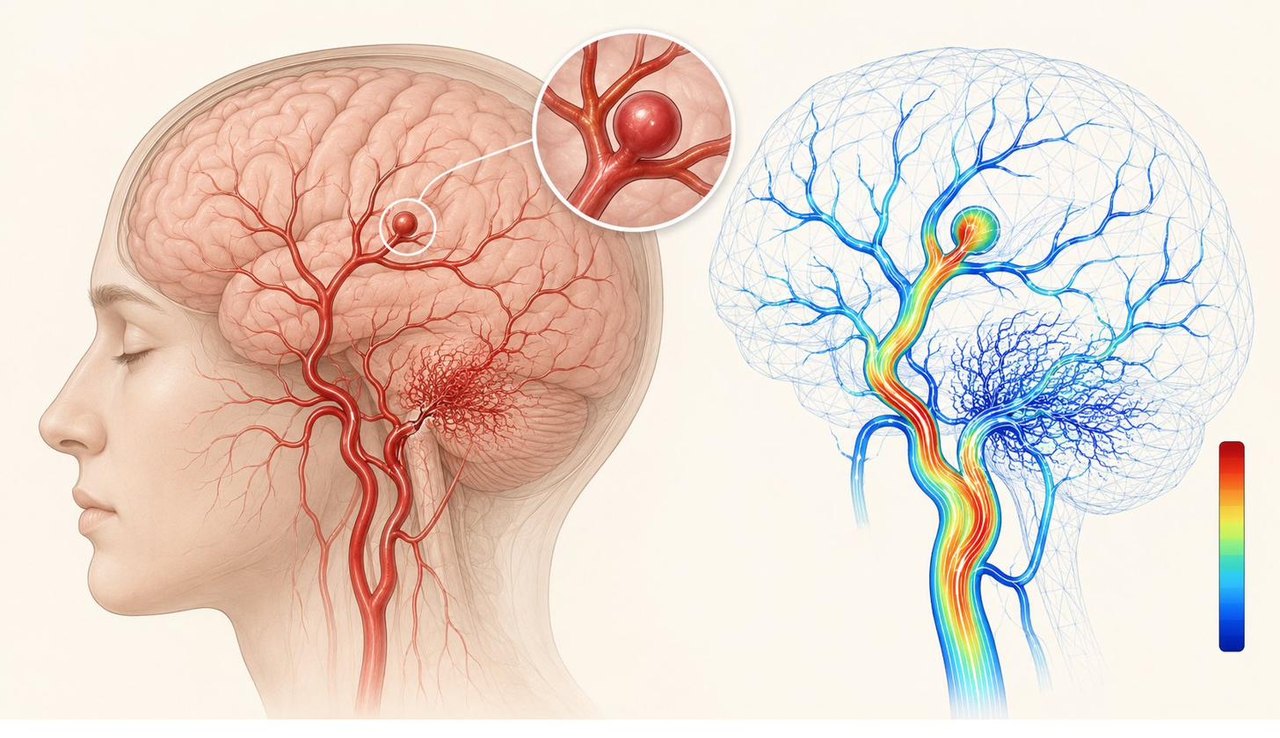
What if a surgeon could see how a patient's brain blood flow will respond to an operation before the operation itself? That is where this theme is headed. The lab studies the fundamental physics of how blood flows and deforms, and turns it into patient-specific digital twins of neurovascular disease such as moyamoya, aneurysms, and stenosis, where a faithful model of an individual's blood flow can guide how neurosurgeons plan a procedure. Building these digital twins draws on theoretical modelling of blood flow, computational fluid dynamics (CFD) of diseased vessels, and experimental microfluidic disease models such as hydrogel-based vessel constructs that reproduce a diseased geometry and test the simulations against real flow. A student can take up any one of these approaches or move between them, so there is a place here whether your strength is analysis, simulation, or the bench.
Click a project title to read its description.
In moyamoya disease, the brain's own arteries slowly close. A fragile web of small vessels grows to compensate, and the surgeon must decide how best to restore flow with a bypass. This project builds a patient-specific digital twin of that circulation, an accurate, image-based model of the patient's vessels in which blood is treated as the non-Newtonian fluid it is, and uses it to predict how a planned bypass will redistribute perfusion across the brain. Microfluidic phantoms of the diseased and rerouted vessels will give the simulation a physical check against real flow, so a bypass plan can be weighed before the operating theatre rather than during it.
An aneurysm is a balloon-like weakening in an artery wall. Clipping it means placing a tiny clip across the neck, shutting the bulge out of the circulation while the healthy branches feeding past it stay open. This project builds a patient-specific digital twin of the aneurysm and its surrounding vessels, an accurate, image-based flow model in which the pulsing wall and the blood are solved together, through fluid-structure interaction, where their coupling matters. With it, a surgeon can test where a clip should sit and see beforehand how each option reshapes the flow and whether the branches stay supplied. Microfluidic phantoms of the patient's vessels will give the simulation a physical check against real flow.
Not every narrowed brain artery is equally dangerous. A stenosis matters when it starves the tissue downstream of blood, and judging that, deciding whether to open the vessel or manage it medically, turns on how much flow the narrowing actually costs. This project builds a patient-specific digital twin of the stenosed artery, an accurate, image-based flow model that resolves the pressure drop across the narrowing and the perfusion left for the brain beyond it, solving the compliant wall and the blood together through fluid-structure interaction where the narrowing's give shapes the flow.
Microfluidics, Biosensing, and Point-of-Care Medical Devices
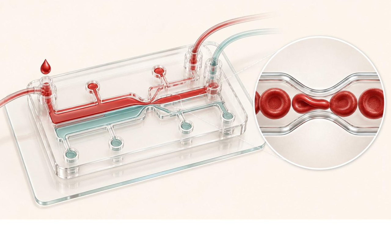
Imagine shrinking a hospital pathology lab onto a chip you can hold in your hand. That is the heart of this theme: portable microfluidic devices and biosensors that read biomarkers from a single drop of blood or body fluid, so a test that once needed a central laboratory can run at the bedside, in a clinic, or out in the field. You might design the chip, build the biosensor, develop the assay, or decode the signal, and students from the life sciences, chemistry, pharmacy, and engineering each find a natural way in. The work is hands-on and tangible, and you need no modelling or coding background to begin.
Click a project title to read its description.
In conditions such as pre-eclampsia and anemia, the physical behaviour of blood shifts in ways a routine blood test was never built to see. This project develops a handheld microfluidic device that reads two of those physical signatures, the viscosity of whole blood and how readily red cells deform, from a single drop, bringing a measurement that today needs a hospital laboratory to the point of care. Its first clinical aim is to grade the severity of pre-eclampsia, with anemia screening as a parallel target.
Sepsis quietly reshapes the blood: red cells stiffen, they clump together, and the blood as a whole begins to flow differently. This project works out the physics of that change, from how blood cells alter their mechanics in sepsis to the flow signatures those changes leave in a microchannel, and it does so through theory and computational modelling of cellular and whole-blood behaviour. The understanding it builds is the engine beneath the point-of-care sepsis triage device, telling us which physical signals are worth measuring and why.
In sepsis, hours matter, yet the test that confirms it usually sits in a central laboratory the patient cannot reach in time. This project builds a low-cost microfluidic chip that turns the flow signatures of sepsis-altered blood, the very signals the modelling work identifies, into a reading that can be taken at the bedside or in a small clinic. The aim is an early triage tool that flags who needs urgent care, built simply enough to work where a laboratory is far away.
Blood does not flow the same in everyone. Its thickness and the way it moves shift with how many cells it carries and what the plasma holds, and this project studies that dependence directly, mapping how the composition of blood sets its viscosity and flow behaviour through controlled experiments and the physics that explains them. It is the groundwork beneath the device projects, since a chip can read a physical signal reliably only once we understand what shapes that signal in the first place.
Microplastics, Biofilms, and Microbial Flows
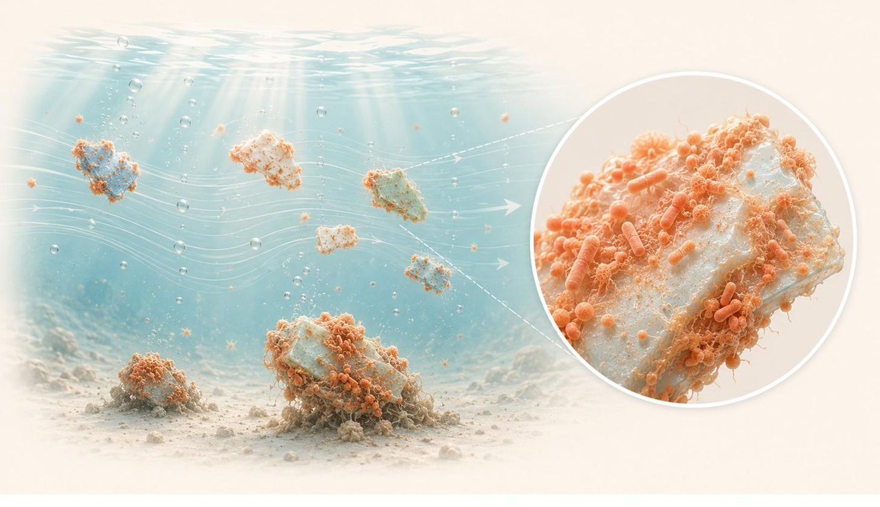
The lab studies how microbes, biofilms, and microplastics move and interact in flows, and what that means for human health and the environment. Questions include how shear flow shapes biofilm growth, how pathogens travel at the micro-scale, how microplastics carry biofilms into the body, and how microbially induced calcite precipitation (MICP) can be harnessed. The work runs from fundamental transport physics to health and environmental impact and combines experiment and simulation, suiting students from biology, environmental science, and engineering.
Click a project title to read its description.
Microplastics rarely travel alone. In water and in the body, microbes settle on them and grow into biofilms, and that living coat changes how the particles move, how they sink or stay suspended, clump, and drift. This project studies the hydrodynamics of these biofilm-coupled microplastics, combining experiment and simulation to follow how the coupling reshapes their transport. The questions reach in two directions at once: where these particles end up in the environment, and how they might ferry microbes into the human body.