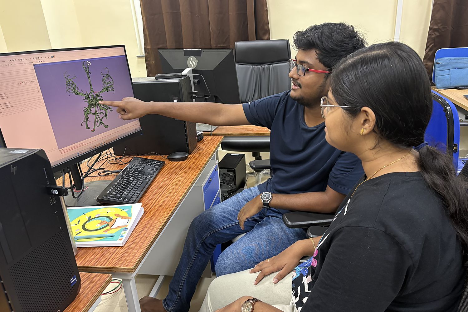
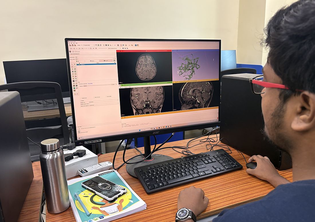
Talking through a cerebral arterial tree reconstructed from an MR angiogram.Segmenting the cerebral vasculature in 3D Slicer. The axial, coronal and sagittal angiography views on the left drive the vessel geometry on the right.
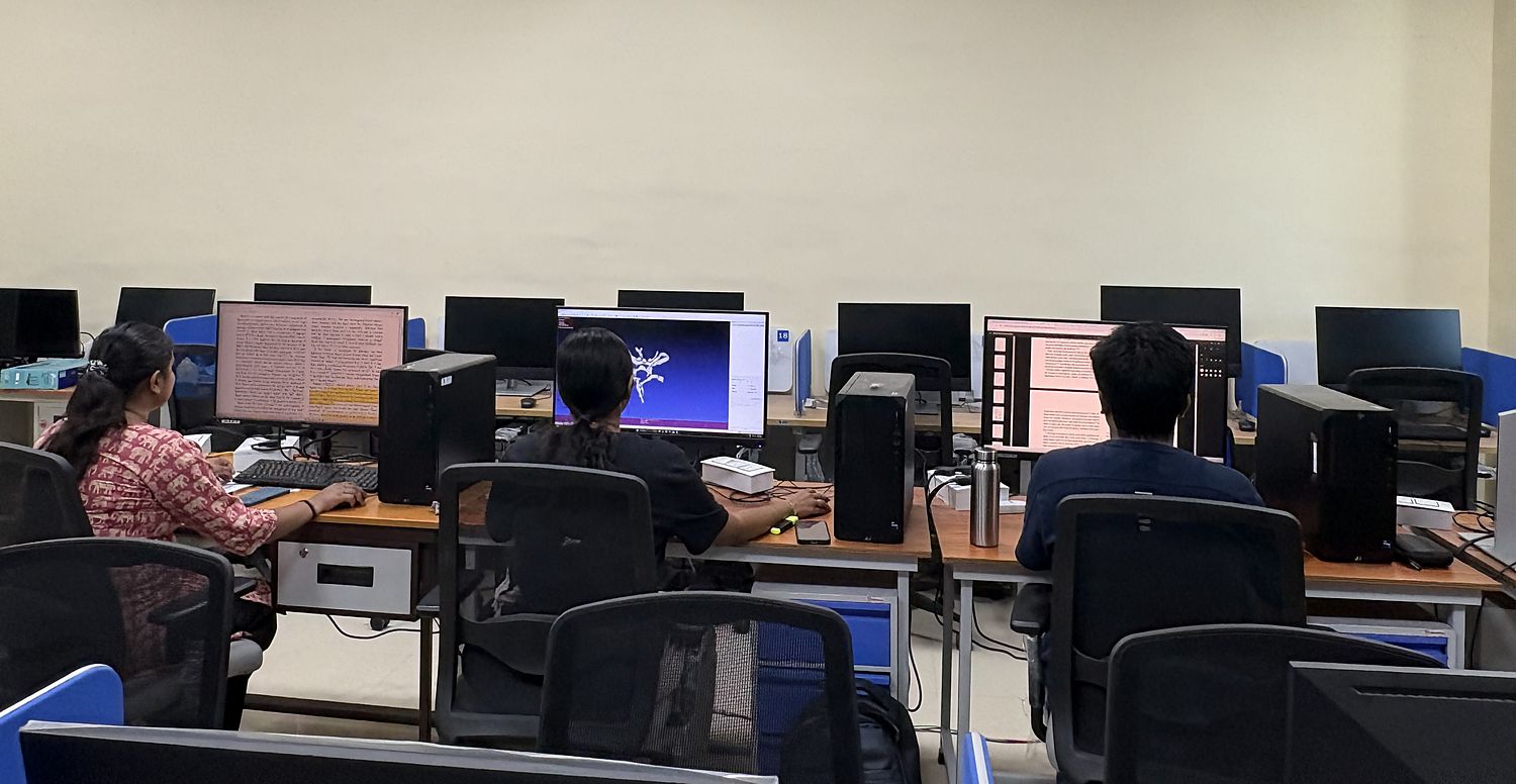
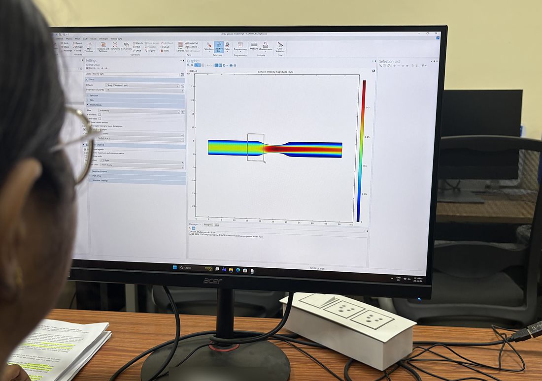
A working morning in the computing room.Velocity magnitude for a Carreau-Yasuda blood model flowing through a constricted vessel, computed in COMSOL Multiphysics.
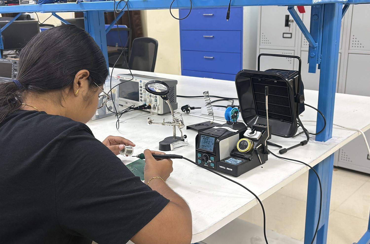
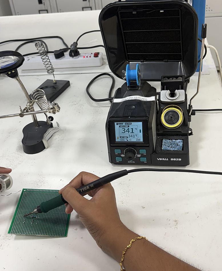
At the soldering station in the prototyping lab, with the bench oscilloscope and the fume extractor alongside.Hand assembly of a prototype board.
At the bench
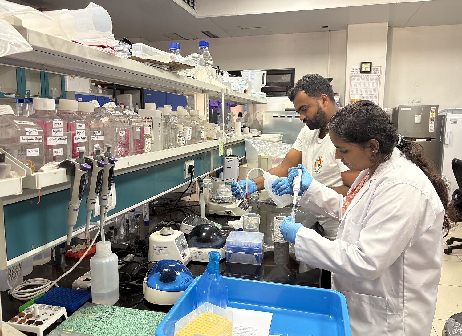
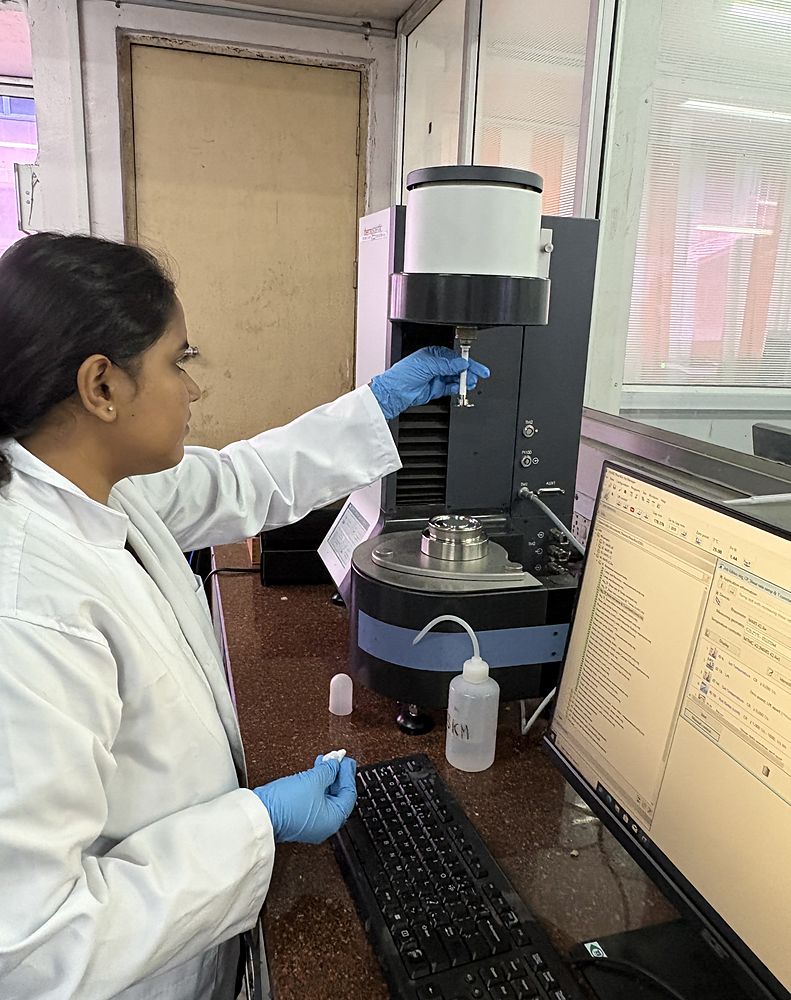
Preparing samples at the wet bench, with the buffer stocks and the microcentrifuges to hand.Mounting the measuring geometry on the rotational rheometer, ahead of a viscosity measurement.
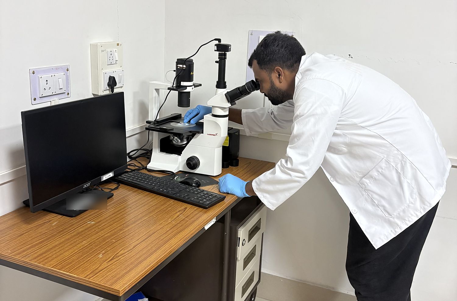
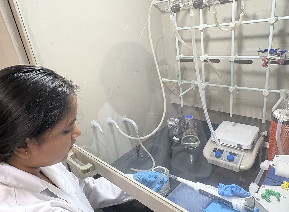
At the inverted microscope, with a sample dish on the stage and the camera trained through the trinocular head.Pipetting inside the fume hood.
At AIIMS Guwahati
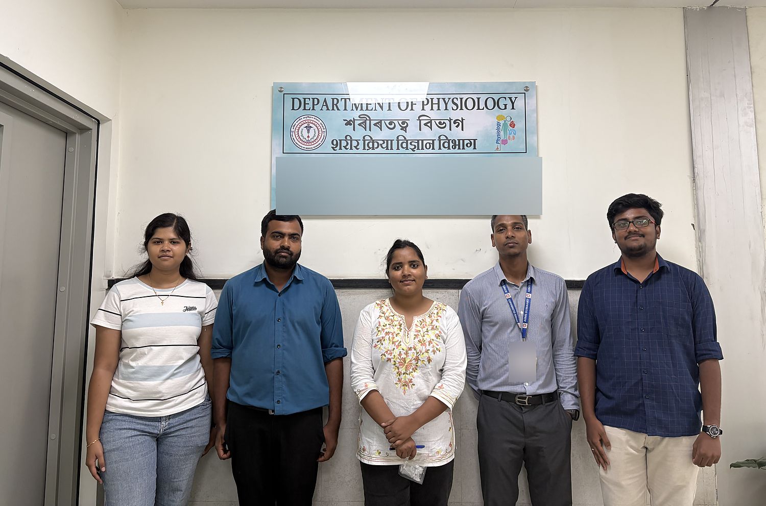
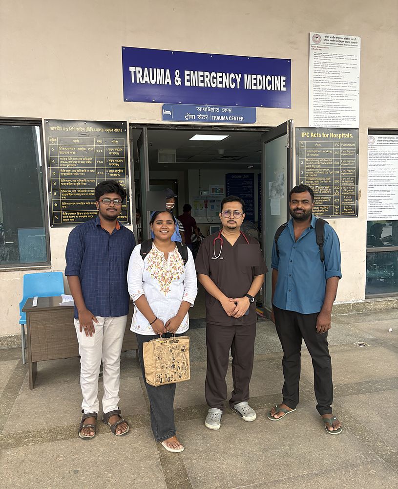
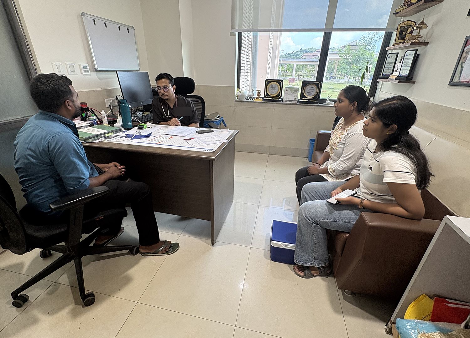
Members of the lab at the Department of Physiology, AIIMS Guwahati, where part of the clinical work is carried out.At the entrance to the Department of Trauma and Emergency Medicine, AIIMS Guwahati.
Working through the clinical side of the study with the Department of Trauma and Emergency Medicine.
The HT5004 class
September 2026
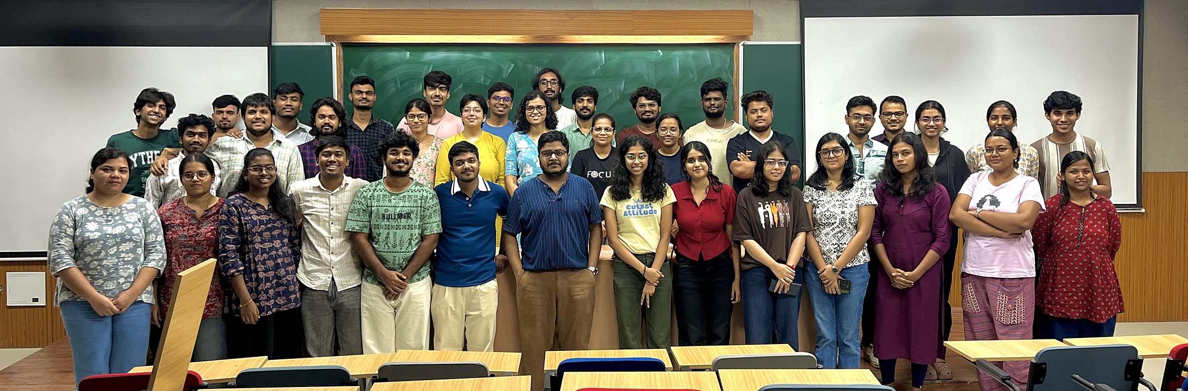
The HT5004 (Basics of Mathematical Modelling and Simulation) class, photographed on 7 September 2026 at the close of the portion of the course taught by Dr. Rahul Agarwal.
Teachers' Day
September 2026
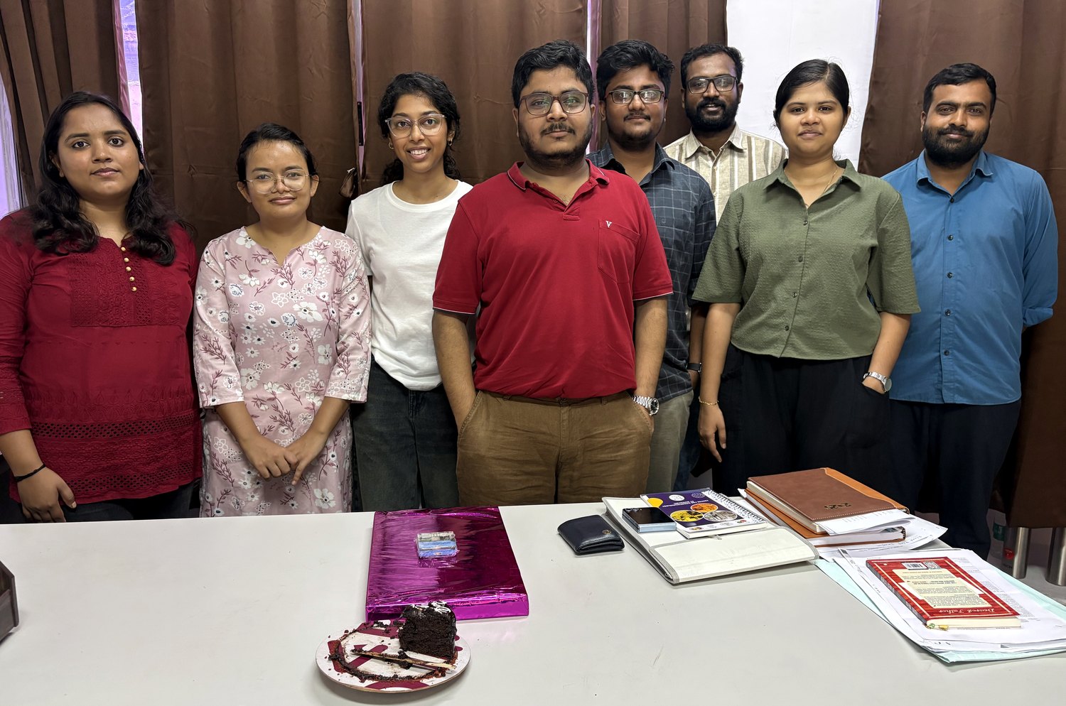
Teachers' Day in the office, 5 September 2026. From left: Jyoti Kumari, Jobila Myrphet, Ananya Jauhari, Dr. Rahul Agarwal, Praanesh C, Rithish Prabhu A, Priyadharshini J M, Dr. Sandeep Kumar Yadav.
India BioImaging training course
June 2026
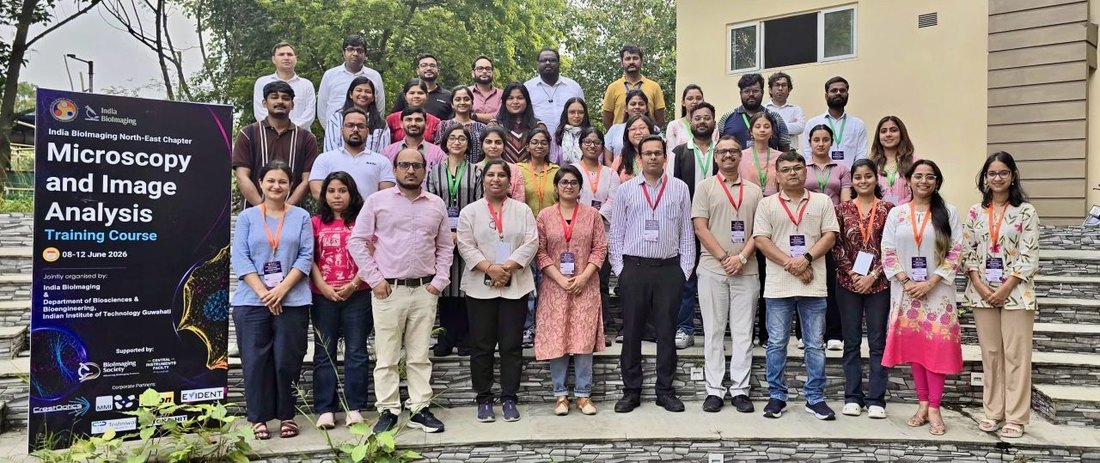
India BioImaging Microscopy and Image Analysis Training Course, IIT Guwahati, 8 to 12 June 2026.
Lab dinner
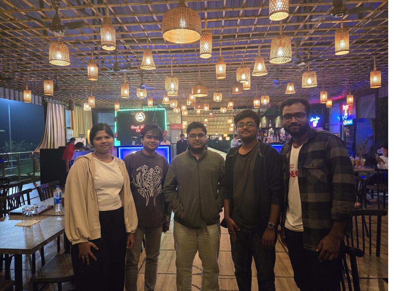
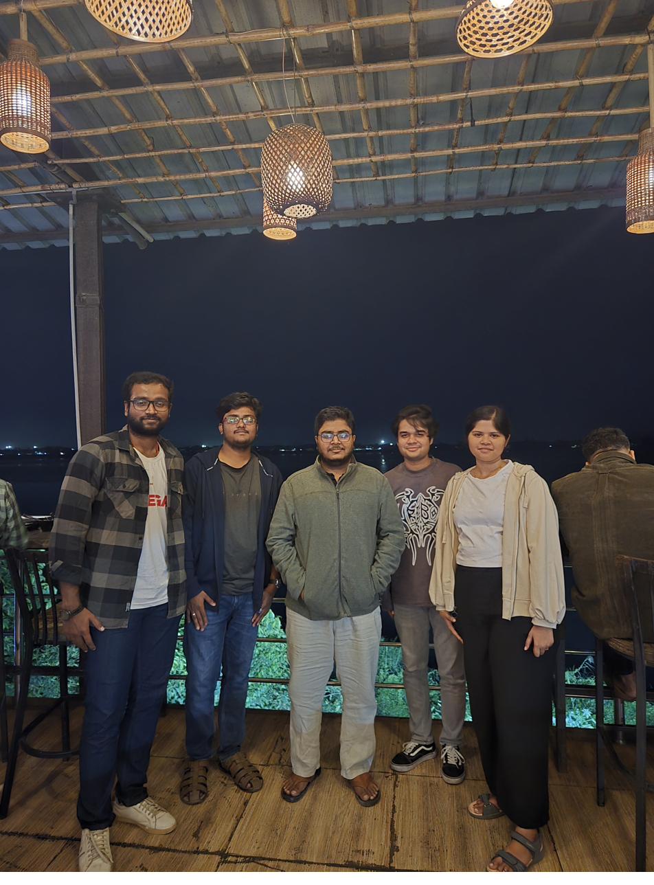
November 2025
From left: Priyadharshini J M, Aditya Meghwal, Dr. Rahul Agarwal, Praanesh C, Rithish Prabhu A.From left: Rithish Prabhu A, Praanesh C, Dr. Rahul Agarwal, Aditya Meghwal, Priyadharshini J M.
Prof. Anupam Sengupta's visit
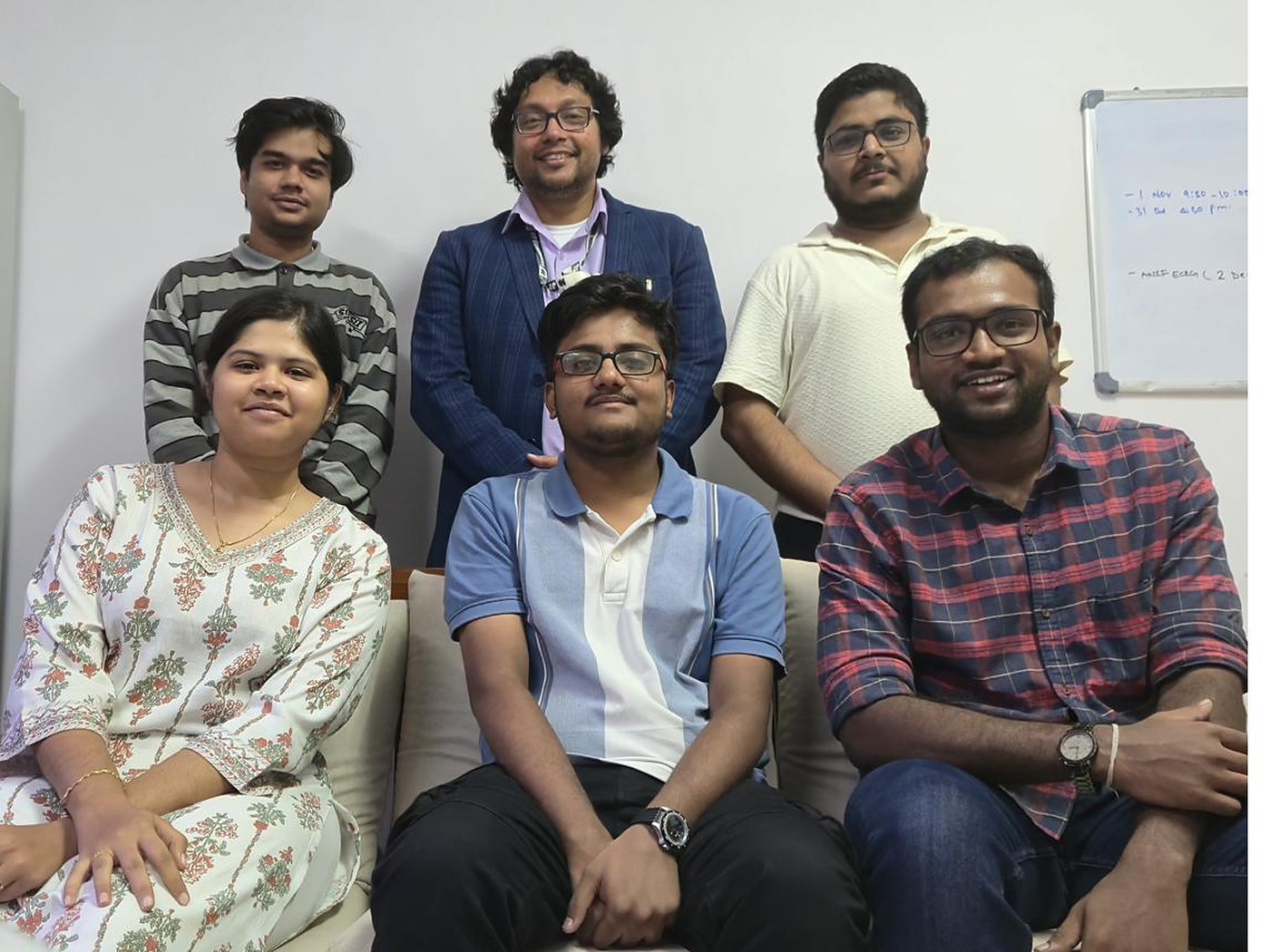
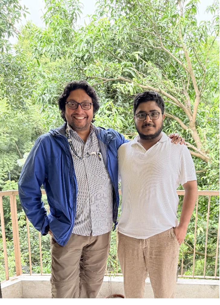
September 2025
In the office. Standing, from left: Aditya Meghwal, Prof. Anupam Sengupta, Dr. Rahul Agarwal. Seated, from left: Priyadharshini J M, Praanesh C, Rithish Prabhu A.At home. From left: Prof. Anupam Sengupta, Dr. Rahul Agarwal.
 Biosensing & Biophysics Lab